
Advanced Cardiac Imaging
- Faculty
- Echocardiography
- Cardiac CT
- Cardiac MR
- Nuclear Cardiology
- Clinical Trials
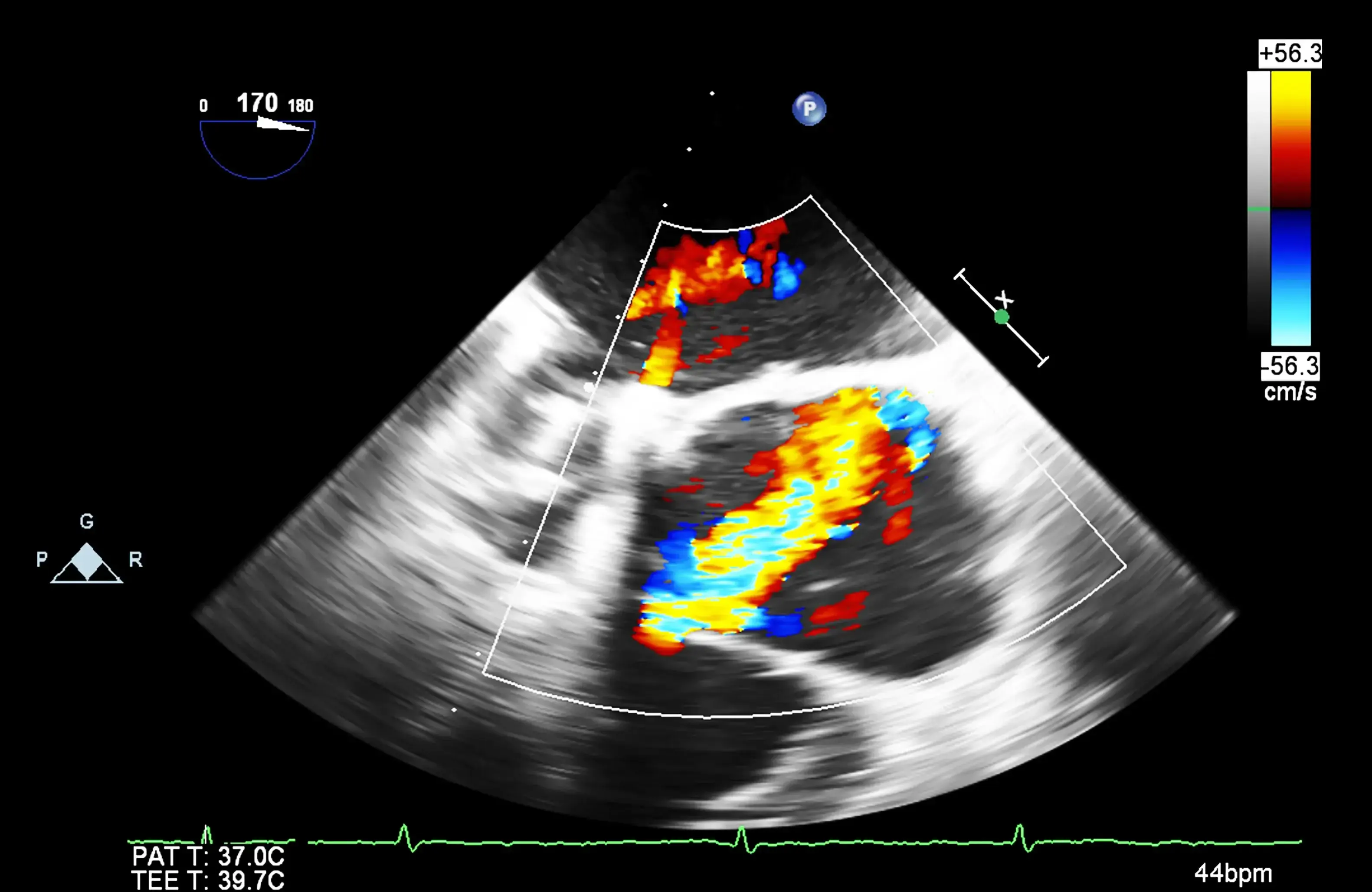 An echocardiogram (echo) uses sound waves to produce moving pictures of the heart chambers, heart valves, and the blood flow through them. Along with the electrocardiogram, echo is one of the basic tests used to evaluate the heart in almost all patients.
Echo is a non-invasive technique in which a small sensor is placed on the chest and directed toward different heart structures. It is painless, safe, and does not require radiation. The images which are obtained can be used to determine the heart's structure, size, and function, including the condition of the heart valves, the thickness of the heart walls, and the velocity and turbulence of blood flow.
Echos can be used to diagnose and monitor many heart disorders. It is of value in diagnosing heart failure, heart valve malfunction, defects occurring at birth, inflammation and infection of the heart or surrounding tissue, and abnormalities of the major vessels that carry blood flow from the heart. In patients with disease of the coronary arteries, it can detect inadequate blood flow during exercise or stimulation and abnormal pumping after a heart attack.