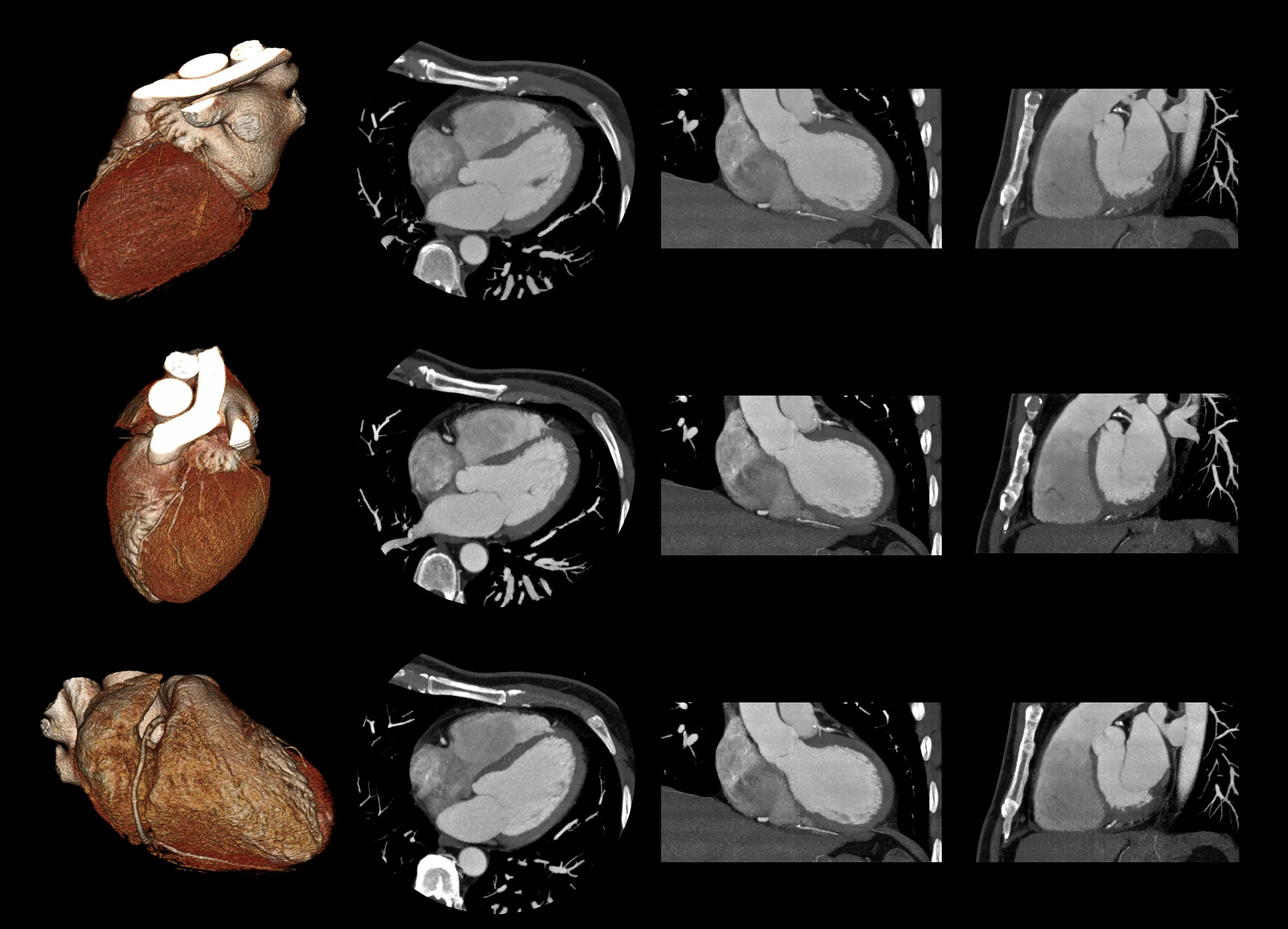
Cardiac CT
 Cardiac CT (computed tomography) is a medical imaging technique that uses computerized processing of X-rays to construct detailed images of the heart, the major blood vessels with which it is attached, and the coronary arteries that bring blood to the heart muscle itself. It is safe and painless and involves exposure to only a very small amount of radiation. The procedure takes less than an hour.
Cardiac CT may be used without administration of a contrast agent to detect calcium in the walls of the coronary arteries, or with an intravenous injection of x-ray contrast agent to visualize blood flow through the channels of the coronary arteries. It is the only technique that enables direct visualization of plaques within the coronary arteries that can lead to heart attacks. It can also provide high-resolution images of the heart's structure and function.
At UCSD our 320-slice CT scanner can complete a whole-heart coverage scan in less time than a heartbeat, providing quality evaluations while minimizing patient exposure to radiation.
